The majority of researchers believe two proteins are responsible for the Alzheimer's disease: tau and beta-amyloid.
Alzheimer’s has notoriously been one of the most difficult diseases to battle. Aging increases the risk of developing the disorder and there is no way to stop the aging process.
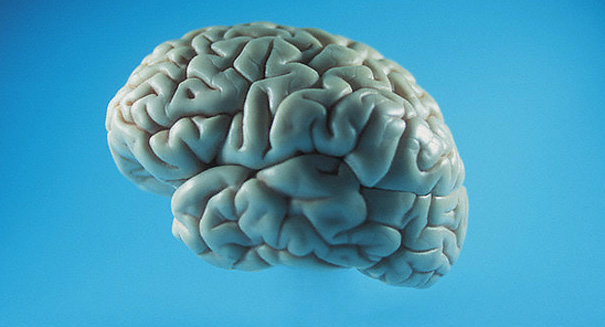
The majority of researchers believe two proteins are responsible for the Alzheimer’s disease: tau and beta-amyloid. It is believed that as the aging process takes place, these two proteins disrupt signaling between neurons or kill them altogether. However, a new study from UCLA suggest a new possibility for the cause: accumulation of iron.
A professor of psychiatry at the Semel Institute for Neuroscience and Human Behavior as UCLA and senior author of the study, Dr. George Bartzokis and his colleagues analyzed two areas of the brain in patients diagnosed with Alzheimer’s. They compared the hippocampus, known to receive damage in the early phases of the diseases, and the thalamus, an area typically not affected until the later stages of the disease. Through the use of sophisticated brain imaging technology, it was discovered that iron is increased in the hippocampus and associated with tissue damage in the area. However, no increase in iron was found in the thalamus during analysis.
Most Alzheimer’s research in the past has focused on the buildup of tau and beta-amyloid in signature plaques associated with the disease. Dr. Bartzokis, however, has long believed that the breakdown begins much further upstream. He says the destruction of myelin, fatty tissue coating the nerve fibers in the brain, disrupts communications between neurons and promotes that buildup of plaques. As a result, the amyloid plaques destroy more myelin, which disrupts signals in the brain, leads to cell death and presents the classic clinical signs of the disease.
In this new study, Dr. Bartzokis and his colleagues tested their hypothesis that levels of iron caused the tissue to break down. An MRI technique that can measure the amount of brain iron in ferritin, a protein that stores iron, was used to analyze 31 patients with the disease and 68 healthy control group participants.
“It is difficult to measure iron in tissue when the tissue is already damaged,” Bartzokis said. “But the MRI technology we used in this study allowed us to determine that the increase in iron is occurring together with the tissue damage. We found that the amount of iron is increased in the hippocampus and is associated with tissue damage in patients with Alzheimer’s but not in the healthy older individuals or in the thalamus. So the results suggest that iron accumulation may indeed contribute to the cause of Alzheimer’s disease.”
“The accumulation of iron in the brain may be influenced by modifying environmental factors, such as how much red meat and iron dietary supplements we consume and, in women, having hysterectomies before menopause,” he added.
This research can be read in the August edition of the Journal of Alzheimer’s Disease.
Leave a Reply