The pictures look like something you might see in a biology textbook, rich and detailed photos of tiny microscopic organisms that cannot be glimpsed with the naked eye.
Despite what the modern Instagram world would have you believe, photography isn’t all about pictures of food and rustic color filters. Nikon proved precisely that this week when it announced the winners from its annual Small World Photomicrography Competition. The pictures look like something you might see in a biology textbook, rich and detailed photos of tiny microscopic organisms that cannot be glimpsed with the naked eye.
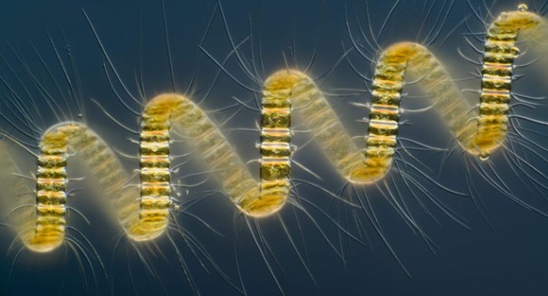
According to an article published on Wednesday by the Los Angeles Times, the winning photo – which depicts a marine diatom colony, a type of golden yellow plankton that lives in colonies and boasts innumerable white bristles – captures a space of just 1/2 millimeter in length. Taken by photographer Wim van Egmond from the Micropolitan Museum (located in the Netherlands), the picture used a 250X magnification in order to capture the visual nuances of the marine diatom. In contrast, van Egmond said that the human eye might be able to pick up on the organism by placing it in a jar of water and holding it up to the light, but that the visual would be little more than a tiny dot to the naked eye.
Still, van Egmond didn’t exactly have the most state-of-the-art equipment on his hands when he went to take the picture that won him the Nikon contest. In fact, the photographer was working with a decrepit light microscope from the 1970s. He also used a glass slide and a thin plastic cover to capture the image in highest resolution possible. Sound familiar? For those who took a biology class in high school or college, the answer is probably “yes.”
With that said, van Egmond isn’t entirely living in the past. While his microscope set-up may not be what a full-time scientist’s would, his photography skills are undeniably on display in the photograph that he entered into the Small World Photomicography Competition. Instead of taking several dozen photographs and choosing the best one for the contest, van Egmond took pictures of different segments of the organism and then built them into a single composite photograph with a computer program. It’s a testament to the photographer’s artistic skill that the final product is completely seamless.
Nikon’s Photomicography Competition is open to anyone “with an interest in microscopy and photography.” However, many of the high-placing entrants are scientists due to the resources they possess for taking microscopic images. Most of van Egmond’s closest competitors were scientists from noted research facilities.
See the rest of the winners here.
Photo credit: Wim van Egmond
Leave a Reply